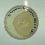
Discovery of Noonan
Collectors Name: Amanda Stone
Date of collection: September 1, 2017
Sample type: Moist Soil
Location Description: An old fox hole
General location: Glen Rose, Texas
Specific location: Latitude 32.23473808 Longitude -97.76084532 Altitude: 175
Sample Depth: 11 cm
Ambient Temperature 90 degrees Fahrenheit
Sample 2
Date of collection: September 1, 2017
Collectors Name: Amanda Stone
Sample Type: Dried out dirt
Location Description: Compost pile
General location: Glen Rose, Texas
Specific Location: Latitude: 32.23675776 Longitude -97.6047711 Altitude 170.0
Sample Depth: 7 cm
Ambient Temperature: 90 degrees Fahrenheit
Sample 3
Collectors name: Amanda Stone
Date of collection: September 3, 2017
Sample Type: Wet soil
Location Description: Grassy area
General Location: Stephenville, Texas
Specific Location: Latitude: 32.21467232 Longitude: -98.20205101 Altitude: 381
Sample depth: 6 cm
Ambient temperature: 91 degrees Fahrenheit
9/6/2017
Following protocol I began the process of making the enriched sample. The protocol called for the use of a 15 ml conical tube, but instead the whole 50 ml conical tube that was used for the collection of the sample was used in the process. The protocol calls for the conical tube to be placed into a shaking incubator for a minimum of an hour. I allowed my sample to incubate from 10:33 a.m.- 12:24 p.m. While my direct sample was in the shaking incubator from 10:30- 11:30 a.m. After tthe direct sample incubated 1.5 ml of the liquid sample was taken from the conical tube and placed into a microcentrifuge tube. Further following the protocol the sample was then put onto a plate. My plate had 2 small bubbles near the wall of the plate.
9/11/2017
1 ml of the enriched sample was placed into a microcentrifuge tube and approximately .8 ml of the enriched sample was placed into another, the two samples were then centrifuged for one minute. After the samples centrifuged for one minute they were filtered and prepared for the spot test following protocol in the manual. Before the samples were placed onto the plate a top auger that contained host bacteria was placed onto the plate and allowed to solidify for approximately 20 minutes until it had solidified. Once the spot test was prepared following the protocol , the plate was then placed into the incubator at 10:35 a.m. The spot test was then allowed to incubate until 9:13 a.m on September 13, 2017. The spot test contained no visible phage, but contained clear indications of contamination meaning that the aseptic technique was not correctly applied to putting the sample onto the plate. Due to the contaminated plate i had to redo the spot test, before i was able to spread the top auger around the entire plate it had partially solidified forcing me to make yet another plate for the spot test. The new plates auger was allowed to solidify for approximately 20 minutes. The second time the spot test was executed with the double filtered enriched sample the plate was bumped before the samples had an adequate amount of time to seep into the top auger. The spots shifted slightly to the left. The new spot test was allowed to sit undisturbed for approximately 15 minutes allowing the spots to adequately seep into the top auger. The spot test was then placed into the incubator at 10:40 a.m. on September 13, 2017. Due to no phage being discovered in my samples i was forced to adopt a sample from a fellow classmate. I adopted my phage from Esperanza Sandoval. The following is information she recorded about the sample in which her phage was discovered.
- Collector name :Esperanza and Raul Sandoval
- Date of collection 09-02-2017
- Sample Type: Soil
- Location Description: A ranch, in a picadero, where horses were being trained to compete.
- Specific location: N 32 40′ 47.81746″ Longitude W 9715’11.14521″
- Sample Depth: one inch
- Ambient temperature 87 degrees Fahrenheit
After adopting a phage i moved onto protocol 6.2 Serial Dilution. Before starting the dilutions i prepared the plates for each dilution and allowed the top auger to solidify as i did the dilutions following protocol 6.2. After each dulution was placed onto plates i placed the dilutions into the incubator at 10:50 a.m on 9/20/17. The first set of serial dilutions were removed from the incubator on 9/22/17 at 9:26 a.m. Taking a phage from the first dilution set i followed protocol 6.2 to prepare a second serial dilution plate set. The second dilution set was placed into the incubator at 10:45 a.m. on 9/25/17 and removed at 9:26 a.m. on 9/27/17. Folowing protocol i flooded a plate and placed into a 4 degree celsius fridge at 10 a.m. on 9/27/17 and removed it at 4 p.m the next day. I then performed a lysate dilution and spot titer test following protocols 6.2 and 6.4 of the manual. The spot titer test was allowed to seep into the top auger for approximately 25 minutes and then placed into the incubator at 5:10 p.m. on 9/28/17. Due to the plate being in the incubator for too long it dried up and was unusable. I then was forced to perform another spot titer test following the same protocol previously stated. this spot titer test incubated from 10 a.m on 10/6 to 9 am on 10/11. The spot titer test did not correctly show making me have to do yet another spot titer test. The third attempt was yet another incorrect showing and was partially dried. It incubated for a little over 24 hours. The next spot titer i plan to only allow to incubate for 12 hours before it is checked to better be able to see the growth of my phage progress.
