Discovery of Puggeroni
Sample Collection
The sample was collected on September 3, 2018 from a ranch in Stephenville, Texas (32° 19° 37° N, 98° 11° 22° W). Recorded temperature at the time was 80° F (27° C) .
September 5, 2018.
Dylan and I adopted a Phage from Matt Bristerpostma who conducted the first few steps of the Virus Isolation process. Matt completed a direct isolation to isolate this phage.
Direct Isolation
This method extracts phages from microbes and particulate matter in a solid or liquid environmental sample. The extracted sample is then used to infect your host bacteria by using the plaque assay. This method offers a snapshot of all the phages present in your environmental sample that may infect your host.
Procedure
- Using a 15 ml conical tube, fill it approximately one-third to one-half full with soil.
- Add liquid media until the sample is submerged beneath 2–3 ml of liquid.
- Cap the tube and invert several times to mix thoroughly.
- Incubate the tube while shaking vigorously in a shaking incubator at 250 rpm for 1–2 hours.
- Allow the sample to sit until particulate matter has mostly settled. Anywhere from 2- 20 minutes.
- Prepare a phage filtrate using aseptic technique.
- Open the package of a syringe filter (0.22 μm), leaving the filter in the packaging.
- Using a syringe, remove approximately 2 ml of liquid from the top of the flooded sample.
- Avoid withdrawing any solid material from the bottom of the tube to prevent clogging the filter during filtration.
- Attach the syringe to the top of the filter, and then remove the filter from the package. (Be careful not to contaminate the filter in the process).
- Make sure the filter is screwed firmly into place.
- Depressing the syringe plunger, dispense a minimum of 0.5 ml of filtrate into a labeled microcentrifuge tube.
- Because debris can clog the filter, you may encounter resistance
- Do not continue to force liquid through the filter or it will break.
- If your filter clogs, remove the clogged filter, replace it with a new one, and continue filtering
- Cap the tube immediately.
- Discard the syringe and filter.
- Proceed to Plaque Assay
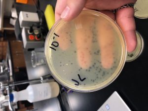
September 7th, 2018.
Plaque Assay, following Direct Isolation.
Overview from Phage Discovery Guide, Section 5.3 : The plaque assay allows you to visually confirm the presence of phage particles in a sample. It is a versatile assay that can be used for phage isolation, purification, and titering. In a plaque assay, host bacteria are mixed with a phage sample and grown as a lawn on agar. If phages are present, they will infect and replicate within the bacterial host, killing the host in the process. Newly replicated phages diffuse within the agar and repeat the process of infection, replication, and lysis of nearby host bacteria. As a result, a visible circular zone of clearing/killing called a plaque will become apparent on the bacterial lawn. Note that each plaque arises from a single phage particle in the original phage sample.
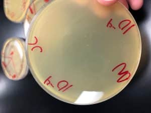
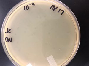
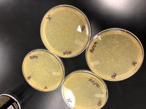
Following 24 hours there was a mostly clear plaque.
- Day 1
- Prepare bench with aseptic technique
- Gather samples you want to assay
- Inoculate the host bacteria with your phage sample
- Obtained same amount of aliquots of 250 microliters host bacteria cultures as phage sample, plus one for negative control.
- Label cultures tubes
- Obtained same amount of aliquots of 250 microliters host bacteria cultures as phage sample, plus one for negative control.
- Used micropipetter and aseptic technique to
- Dispense each phage sample into the appropriate culture tube containing 250 microliters of host bacteria (500 microliters)
- Mix inoculated host culture by tapping tube
- Let sit undisturbed for 5-10 minutes for attachment
- Plate samples with top agar
- Obtained plate
- Removed agar from 55°C bath
- For each sample
- Using a sterile 5 mL pipette, aseptically transfer 3 ml of top agar to an inoculated host tube
- Immediately suck up the mixture back into pipette and transfer it to the plate
- Tilt the plate in multiple directions until the top agar mixture evenly coats the agar plate
- Repeat for each plate
- Incubate plates to allow bacterial growth and phage infection
- Let agar settle undisturbed for approximately 20 minutes
- After the top agar solidifies, gently invert plate and place in incubator
- Incubate the plates at specified temperature for 24-48 hours (29° C)
- Day 2
- Check the plate for plaques
- Remove plate from incubator and held up to light to check for plaques
- If no plaques return to incubator
- Record Results
- No photo, need additional data from Matthew Bristerpostma
- Mostly clear plaque
- Remove plate from incubator and held up to light to check for plaques
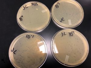
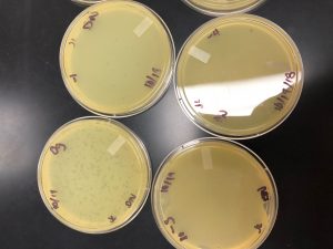
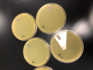
Plaques from Plaque assay done on September 5, 2018

September 10, 2018
Picking a Plaque
From: Phage Discovery Guide, Section 5.4
- Aseptic technique to prepare bench
- Labeled plaque to be picked
- Record Morphology
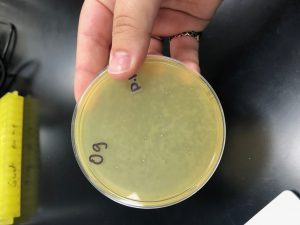
- Mostly clear plaque
- Labeled and prepare microcentrifuge tubes
- As many as plaques picked
- Label each tube
- Using aseptic technique, aliquot 100 μl of phage buffer into each microcentrifuge tube.
- “Pick a plaque”
- Place a sterile tip onto a p200 micropipettor.
- Holding the pipettor perpendicular to the agar surface, gently stab the top agar in the center of the plaque .
- Avoid touching the bacteria surrounding the plaque.
- Place the end of the tip into the phage buffer in the corresponding microcentrifuge tube. Then tap the tip on the wall of the tube and pipette up and down to dislodge phage particles. Discard the tip.
- Mix well by vortexing.
- Repeat Steps 1–4 for each plaque you are picking.
September 17, 2018
Serial Dilutions
Matthew Bristerpostma completed the first round of Serial Dilutions on September 17,2018. The purpose of Serial Dilutions is to manipulate the number of phages in a sample.We use serial dilutions to purify, amplify, and titer your phage. This protocol uses 10-fold serial dilutions, meaning that the concentration of phage in each tube is 10 times less than the previous tube, allowing for easy mathematical calculations.
- Set up 10-fold serial dilutions.
- Arranged the proper number of microcentrifuge tubes in a rack and label them 10-1
- Add 90 μl of phage buffer to each of the tubes.
- Perform the 10-fold serial dilution
- Add 10 μl of undiluted phage sample to the “10-1” tube and vortex well.
- The solution in this “10-1” tube contains 1/10th the number of phage particles in the undiluted sample. Also referred to as a 1:10 dilution.
- Use a clean pipette tip for each transfer , vortexing sample well before making each dilution.
- Transfer 10 μl of the “10 -1” sample to the “10-2” tube and vortex well.
- This solution contains 1/100th as many phage particles as your undiluted sample. It can also be referred to as your 1:100 dilution.
- Continue each successive dilution until last tube.
- Add 10 μl of undiluted phage sample to the “10-1” tube and vortex well.
September 19, 2018
Plaque Assay
Matthew Bristerpostma also completed another plaque assay from the serial dilution.
The steps of the plaque assay follow:
Day 1
- Gather samples to assay
- Inoculate host bacteria with phage sample
- 10 microliters of sample from Phage Sample
- 250 microliters of host bacteria
- mix by tapping the tube
- Let samples sit undisturbed for 5-10 minutes for attachment
- Plate samples with top agar
- Incubate plates for growth
- Let sit for 20 minutes for agar to solidify.
- Incubate 24-48 hours
Day 2
- Check plates
- Mostly clear plaques
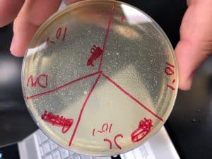
September 26, 2018
2nd Round of Serial Dilutions
Dylan and I completed the 2nd round of Serial Dilutions on September 26, 2018 when we adopted Matthew’s phage. Matthew only had visible plaques up to 10-4 so Dylan and I did dilutions to the 10-6 . We let the plates incubate for 48 hours, and on September 28,2018, we only had plaques on the first three plates, so Jennifer put the plates back in to incubate over the weekend.
- Set up dilutions
- Arrange the proper number of microcentrifuge tubes in a rack and label them 10-1, 10-2, 10-3,….
- Add 90 μl of phage buffer to each of the tubes.
- Performed 10-fold serial dilutions
- Added 10 μl of undiluted phage sample to the “10-1” tube and vortex well.
- Use a clean pipette tip for each transfer and pipette carefully, vortexing sample well before making each dilution.
- Transfer 10 μl of the “10 -1” sample to the “10-2” tube and vortex well.
- Continue each dilution until last tube.
- Added 10 μl of undiluted phage sample to the “10-1” tube and vortex well.
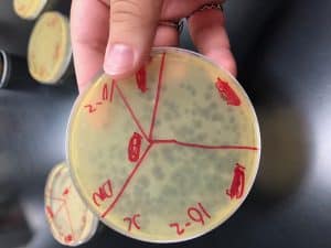
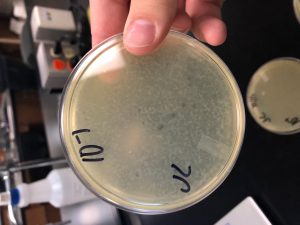
October 1, 2018
Collecting Plate Lysates
We chose our 10-3 plate to collect a lysate from. Visible plaques were only visible up to the 10-3 dilution.
- Flood the webbed plate
- Apply 8 ml of sterile phage buffer to the plate.
- Let the plate sit at room temperature for 3 hours
- Swirl the phage buffer
- Harvested plate lysate.
- Remove the lid from the plate and place it on the bench.
- Tilt the plate slightly by placing one edge of the plate on the lid, allowing the lysate to pool to one side
- Prepare a 0.22 μm filter by opening the packaging but not removing the filter.
- Aspirate the lysate from the plate using 5 mL syringe
- Carefully attach the syringe to the filter. Depress the syringe plunger and collect the filtrate in a 15 ml sterile conical tube.
- Label tube
- Storing lysate at 4 ° C
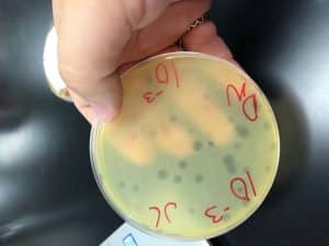
October 8, 2018
Spot Titer with Serial Dilutions from Collected Plate Lysate
- Label one agar plate for a spot titer.
- Labeled the bottom of an agar plate with your name, the date, and the notation “spot titer.”
- We split the plate into 7 sections, six for our dilutions and one for our negative control.
- Next we prepared our bacterial lawn
- Using a sterile 5 ml pipette,we transferred 3 ml of molten top agar into a 5 ml sterile pipette.
- Next, we transferred the top agar to a tube containing 250 μl of host bacteria, immediately drawing the solution back into the pipette.
- Dispensed the top agar-bacteria mixture onto an agar plate, spread evenly around plate and let sit 20 minutes to settle.
-
Serial Dilutions
- Prepared materials with aseptic technique
- Completed serial dilutions to the 10-6
- Added 90 μl of phage buffer to each of the tubes
- Add 10 μl of original phage sample to “10 -1”
- Transfer 10 μl of the “10 -1” sample to the 10 -2 tube and vortex well.
- Continue each dilution until last tube.
-
Spot Titer
- We spotted the dilutions and control on the prepared bacterial lawn plate.
- One at a time, aseptically transferred 3 μl of all samples onto the proper location on the bacterial lawn.
- Use 3 μl of sterile phage buffer as the negative control.
- Allowed the liquid from the spots to absorb into the agar for 45 minutes
- Incubated plates for 48 hours at 29.5 °C .
- Calculating Spot Plate Titer
- Titer (pfu/ml) = (# pfu/ volume used in μl) x (103 μl/ml) x dilution factor
- Found 6 plaques on 10 -4
- (6 pfu/3 microliters) * (10 3 ( “10 -4)
- 2 * 10 7
- 20,000,000
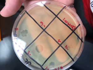
plaques visible up to the 4th dilution
October 10, 2018
Full Plate Titer
- Performed serial dilutions to 10-6
- Add 90 μl of phage buffer to each of the tubes.
- Performed 10-fold serial dilutions
- Added 10 μl phage sample to the “10-1” tube and vortex well.
- Transfer 10 μl of the “10 -1” sample to the “10-2” tube and vortex well.
- Continue each successive dilution until you get to your last tube.
- Complete Plaque Assay to find Full Plate Titer
- Inoculate bacteria with phage sample
- Obtain the same number of aliquots of 250 μl host bacterial cultures as you have phage samples
- Use a micropipettor and aseptic technique to
- Dispense each 10 microliter phage sample into the appropriate culture tube containing 250 μl of host bacteria
- Mix each inoculated host culture by gently tapping the tube.
- Let your sample sit undisturbed for 5–10 minutes to allow for attachment
- We did not let the sample sit for 5-10 minutes**
- Plate the samples with top agar. For this part of the experiment you will need 3 ml of molten top agar per sample. Your instructor may provide this for you or you may need to make it according to the protocol found in the
- Obtain the same number of agar plates as you have samples.
- Using a sterile 5 ml pipette, aseptically transfer 3 ml of top agar to an inoculated host tube
- Immediately aspirate the mixture back into the pipette and transfer it to the appropriate plate
- Gently, but quickly, tilt the plate in multiple directions until the top agar mixture evenly coats the agar plate.
- Repeat this process for each of your samples.
- Incubate plates to allow bacterial growth and phage infection.
- Let the plates sit undisturbed for ~20 minutes until the top agar solidifies.
- Incubate the plates at the specified temperature for 24–48 hours.
- After 48 hours we experienced growth on the 1st dilution plate and we let the plates grow over the weekend.
-
October 15, 2018
- We experienced growth up to our 4th serial dilution.
- No growth on 5th or 6th plate
- Obtain the same number of agar plates as you have samples.
- Use a micropipettor and aseptic technique to


October 15, 2018
Collecting Plate Lysate
- Prepared lab table for aseptic technique
- Collected plate for lysate collection
- We used a plate from our full plate titer. We used our 10-2 dilution
- We counted 70 plaques, and calculated a titer of 7 x (105)
- 700,000
- We flooded the webbed plate, following the protocol
- Apply 8 ml of sterile phage buffer to the webbed plate.
- Let the plate sit at room temperature for 2–4 hours
- Swirl the phage buffer gently, taking care not to splash.
- Collecting a plate lysate.
- Removed the lid and plate
- Prepare a 0.22 μm filter by opening the packaging but not removing the filter.
- Using a 5 ml syringe aspirate the lysate from the plate.
- Attach the syringe to the filter. Depress the syringe plunger and collect the filtrate in a 15 ml sterile conical tube.
- Labeled the tube
- Record Volume of Lysate
- 4.5 mL
- Redoing Serial Diltuons and full plate
- Store the lysate at 4 °C
October 15, 2018
Redid Full Plate Titer
Reconducted full plate titer to double check results and data.
Plated 4 plates with a 10-1, dilution to check titer
- Prepared bench for aseptic work
- Performed serial dilution to the 10-1,
- Innculated bacteria with phage sample
- Sit 5-10 minutes
- Conducted Plaque Assay
- Plate the samples with Top Agar
- Using a sterile 5 ml pipette, aseptically transfer 3 ml of top agar to an inoculated host tube
- Immediately aspirate (suck-up) the mixture back into the pipette and transfer it to the appropriate plate and discard the pipette.
- Incubate plates to allow bacterial growth and phage infection.
- Let the plates sit undisturbed for ~20 minutes until the top agar solidifies.
- Incubate 24-48 hours
-
Full Plate Results
-
October 17, 2018
-


October 17, 2018
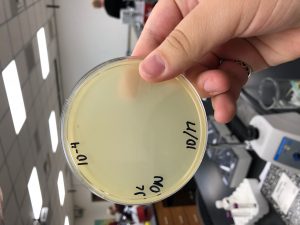
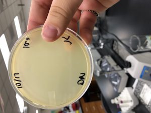
Picking a plaque, Serial Dilutions, and Full Plate
We were receiving different plaque morphology results on our plates from the 10-1, dilution conducted on 10/17/2018. I did
-
Picked Plaque from dilution plate (10/17/2018)
- Small, clear plaque
- Using aseptic technique, aliquot 100 μl of phage buffer into microcentrifuge tube.
- “Pick” a plaque.
- Place sterile tip onto a p200 micropipettor.
- Holding the pipettor perpendicular to the agar surface, gently stab the top agar in the center of the plaque
- Place the end of the tip into the phage buffer in the corresponding microcentrifuge tube. Then tap the tip on the wall of the tube and pipette up and down to dislodge phage particles. Discard the tip.
- Mix well by vortexing.
- Small, clear plaque
-
Serial Dilutions
-
Add 90 μl of phage buffer to each of the tubes.
-
Perform 10-fold serial dilutions
- Add 10 μl of your undiluted phage sample to the “10-1” tube and vortex well.
- Transfer 10 μl of the “10 -1” sample to the “10-2” tube and vortex well.
- Continue each successive dilution until you get to your last tube.
- Full Plate Titer by Plaque Assay
- Inoculate the host bacteria with your phage sample.
- Dispense each phage sample into the appropriate culture tube containing 250 μl of host bacteria
- Mix each inoculated host culture by gently tapping the tube.
- Let your sample sit undisturbed for 5–10 minutes to allow for attachment.
- Plate samples with 3 mL top agar
- Obtain the same number of agar plates as you have samples.
- Remove a bottle of top agar from the 55 °C bath.
- For each sample, including controls using a sterile 5 ml pipette, aseptically transfer 3 ml of top agar to an inoculated host tube
- Immediately aspirate the mixture back into the pipette and transfer it to the appropriate plate and discard the pipette.
- Gently tilt the plate in multiple directions until the top agar mixture evenly coats the agar plate
- Repeat this process for each of your samples.
- Incubate plates to allow bacterial growth and phage infection.
- Let the plates sit undisturbed for ~20 minutes until the top agar solidifies.
- After the top agar has solidified, gently invert the plates and place in the proper incubator.
- Incubate the plates at the specified temperature for 24–48 hours
- Inoculate the host bacteria with your phage sample.
October 19, 2018
Full Plate results from 10/17/2018


October 19,2018
Full Plate Titer- Results from October 17,2018 were invalid. Re-conducting to double check results.
- Used serial dilutions from October 17, 2018
- Inoculated host bacteria with phage sample
- Obtained 7 aliquots of 250 μl host bacterial cultures, six for phage samples, one for negative control
- Used a micropipettor and aseptic technique to dispense each phage sample into tube containing 250 μl of host bacteria.
- Mix each inoculated host culture by gently tapping the tube.
- Let your sample sit undisturbed for 5–10 minutes to allow for attachment.
- Plate the samples with top agar.
- Obtain the same number of agar plates as you have samples.
- For each sample, including control
- Using a sterile 5 ml pipette, aseptically transfer 3 ml of top agar to an inoculated host tube
- Immediately aspirate the mixture back into the pipette and transfer it to the appropriate plate and discard the pipette.
- Gently, but quickly, tilt the plate in multiple directions until the top agar mixture evenly coats the agar plate.
- Repeat this process for each of your samples.
- Incubate plates to allow bacterial growth and phage infection.
- Let the plates sit undisturbed for ~20 minutes until the top agar solidifies.
- After the top agar has solidified, gently invert the plates and place in the proper incubator.
- Incubated the plates for 48 hours.
-
Full Plate TIter Results from 10/19/2018
- Titer: 2.3 x 10⁸


October 22, 2018
Collecting Lysate and Making Webbed Plates
- Apply 8 ml of sterile phage buffer to the webbed plate.
- Let the plate(s) sit at room temperature for 2–4 hours
- Swirl the phage buffer gently, taking care not to splash.
Harvest a plate lysate.- Prepare a 0.22 μm filter by opening the packaging but not removing the filter. Set aside.
- Using a 5 ml syringe aspirate the lysate from the plate.
- Carefully attach the syringe to the filter. Depress the syringe plunger and collect the filtrate in a 15 ml sterile conical tube.
- Label the tube
- Store Lysate
Webbed Plates
Estimate the number of plaques you need and calculate the volume of lysate necessary to generate a webbed plate.
- Using data from a previous plaque assay, estimate how many plaques fill a plate.
- Calculate how many phage were on the webbed plate used to make the lysate.
- Estimate how many more plaques you need to make a webbed plate.
- Calculate the volume of lysate needed to generate a webbed plate.
- 9.2 x 10⁻⁴ μl
- Perform Serial Dilutions
- Plate dilutions
- Infect the bacterial host cells with the appropriate volume and dilutions of your lysate and and incubate the mixture for 5–10 minutes for phage attachment. Then add 3 ml of molten top agar to the mixture and plate.
- Incubate (without inverting) for at least 24 hours at proper temperature.
October 24, 2018
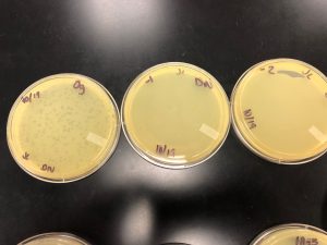
Making Webbed Plates
- Plated 8 plates of undiluted phage (Og) to gather lysate. Phage was only growing to 10-1 dilution,
- Mixed phage with bacteria and let attach for 7 minutes
- Plated sample with top agar
- Added 3 ml top agar to sample and aspirate back up immediately
- Plate mixture, spread quickly and then let sit 20 minutes to solidify .
- Incubate
Day 2: Collecting Plate Lysate
Flood the webbed plate(s).
- Apply 8 ml of sterile phage buffer to the webbed plate.
- Let the plate(s) sit at room temperature for 2–4 hours
- Swirl the phage buffer gently, taking care not to splash.
Harvest a plate lysate.
- Prepare a 0.22 μm filter by opening the packaging but not removing the filter. Set aside.
- Using a 5 ml syringe aspirate (suck up) the lysate from the plate.
- Carefully attach the syringe to the filter. Depress the syringe plunger and collect the filtrate in a 15 ml sterile conical tube.
- Label the tube appropriately.
- Pool the lysates
October 29, 2018
Made Webbed Plates
Plates did not have enough growth on October 26, 2018, so we let the plates sit over the weekend.
- Flood plates with 8 ml of phage buffer
- Let sit for 2-4 hours
- Collect and pool lysate
- 40 ml total

October 31, 2018
Checking titration of lysate by completing Full Plate Titer
- Completed Serial Dilutions
- Add 90 μl of phage buffer to each of the tubes.
Perform 10-fold serial dilutions- Add 10 μl of your undiluted phage sample to the “10-1” tube and vortex well.
- Transfer 10 μl of the “10 -1” sample to the “10-2” tube and vortex well.
- Continue each successive dilution until you get to your last tube.
- Inoculate the host bacteria with your phage sample.
- Obtain the same number of aliquots of 250 μl host bacterial cultures as you have phage samples
- Label the culture tubes accordingly.
- Mix each inoculated host culture by gently tapping the tube.
- Let your sample sit undisturbed for 5–10 minutes to allow for attachment.
- Plate the samples with top agar.
- Obtain the same number of agar plates as you have samples.
- Using a sterile 5 ml pipette, aseptically transfer 3 ml of top agar to an inoculated host tube
- Immediately aspirate (suck-up) the mixture back into the pipette and transfer it to the appropriate plate and discard the pipette.
- Gently, but quickly, tilt the plate in multiple directions until the top agar mixture evenly coats the agar plate.
- Obtain the same number of agar plates as you have samples.
- Repeat this process for each of your samples.
- Incubate plates to allow bacterial growth and phage infection.
- Let the plates sit undisturbed for ~20 minutes until the top agar solidifies.
- After the top agar has solidified, gently invert the plates and place in the proper incubator.
- Incubate the plates at the specified temperature for 24–48 hours.
- Obtain the same number of aliquots of 250 μl host bacterial cultures as you have phage samples
Plate results from Concentration test.
October 31, 2018

November 5, 2018
Microscopy
- Prepare phage samples.
- Aseptically transfer 1 ml of your high-titer lysate into a sterile microcentrifuge tube.
- Balance the tube(s) and centrifuge for 1 hour at 4 °C at top speed to concentrate the phage particles at the bottom of the tube.
- Using a micropipettor, carefully remove as much supernatant as possible without disrupting the concentrated phage at the bottom of the tube.
- Add 100 μl of phage buffer and let resuspend at 4 °C for 30 minutes to one hour.
-
- Put on a fresh pair of gloves.
- Cover the designated work area with bench paper or a Kimwipe to create a clean work surface.
- Remove the cover from a 5 x 5 cm piece of parafilm, and place the parafilm into the lid of Petri dish.
- Place a PELCO Tab or small piece of double-sided tape onto the parafilm in the lid of the Petri dish. Expose the adhesive or the tab.
- Using EM forceps, remove a fresh grid from a box of unused grids, touching only the very edge of the grid.
- Place the grid dark-and-shiny side UP, on the edge of the tab or double-sided tape so that only the very edge of the grid (no more than 0.5 mm) is touching the adhesive.
- Using a micropipettor, place 10 μl of lysate onto the grid without touching the tip to the grid itself.
- Allow the phage settle and attach onto the grid for at least 2 minutes
- Using a small wedge of filter paper, wick off the excess fluid.
- Rinse the grid two times by the following method:
- Carefully pipette 10 μl of sterile water onto the grid. Allow it to sit for 2 minutes.
- Wick off the water using a fresh wedge of filter paper.
- Add 10 μl of 1 % uranyl acetate to the grid.
- Let it sit for 2 minutes.
- Wick off excess stain by using a wedge of filter paper.
- Observe your phage.
- Place grid in the designated grid box for storage.
November 12, 2018
DNA Extraction
Day 1:
- Gently mix your HVL, then aliquot 5mL of your lysate into a 15 mL conical tube.
- Once nuclease has been added, gently invert the tube and incubate at 37°C for 10min.
- Aliquot lysate into 5 microfuge tubes, 1mL each.
- To each tube, add 20uL of ZnCl2, mix gently by inversion, and incubate at 37°C for 5min. This “precipitates” the phage.
- Centrifuge at 10,000rpm for 1min to pellet the phage.
- KEEP THE PELLET. Remove supernatants by aspiration, but try not to disturb the pellet. Discard the liquid filled pipette tips in the sharps trash.
- Resuspend pellets in 500uL TES buffer per tube, and incubate at 60°C for 15min. This will denature the capsids, exposing the DNA, while protecting it from the nuclease activity
- Add 1uL of Proteinase K and mix gently. Incubate at 37°C for 10min to completely eliminate any residual nuclease activity.
- Add 60uL of potassium acetate to each tube. Mix well and leave on ice for 15min. A white, dense precipitate will form. This represents your capsids.
- Centrifuge at 4°C for 1min at 12,000rpm to pellet the capsids. KEEP THE SUPERNATANTS containing your DNA, and place into new microfuge tubes. You can throw away the tubes with the pellets.
- Add 500uL of isopropanol to each of the tubes with the supernatant, mix, and leave on ice overnight

November 14, 2018
Day 2 of DNA Extraction
- Centrifuge at top speed for 10min to pellet DNA, and discard the supernatant into waste tube.
- Add 250uL of 70% ethanol in each tube, and spin again for 1min, at top speed.
- Dry the DNA pellets at room temperature by turning upside-down onto paper towels, tapping out excess liquid, and leaving upside-down until pellets begin to turn clear. The tubes can also be placed in a fume hood or 30⁰C incubator to help with drying.
- Resuspend the first pellet in 50uL nuclease-free water. Then use that solution to resuspend the next pellet. Continue until all 5 pellets have been resuspended in the same 50uL of water.
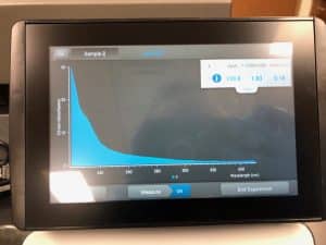
- Check DNA concentration and quality (A260:280 and A260:230) with the Nanodrop.
Results from NanoDrop
ng/ul: 138.8
A260/A280: 1.83
A260/A230: 0.18